Loculated Pleural Effusion / Pulmonology CXRs - Physician Assistant Studies Pa Medicine ... : Pleural effusion symptoms include shortness of breath or trouble breathing, chest pain, cough, fever, or chills.
Dapatkan link
Facebook
X
Pinterest
Email
Aplikasi Lainnya
Loculated Pleural Effusion / Pulmonology CXRs - Physician Assistant Studies Pa Medicine ... : Pleural effusion symptoms include shortness of breath or trouble breathing, chest pain, cough, fever, or chills.. Pleural effusions result from abnormal buildup of a thin layer of liquid that normally helps adhere and lubricate the interface between visceral and parietal pleura. Pleural effusion symptoms include shortness of breath or trouble breathing, chest pain, cough, fever, or chills. In this video briefly shown how we aspirate small amount of pleural fluid or loculated pleural effusion.for more videos please subscribe the channel.if you. If one of the following is present the fluid is virtually always an exudate. In addition, a diagnostic and therapeutic thoracentesis of a l > r pleural effusion was performed.
Loculated effusions are collections of fluid trapped by pleural adhesions or within pulmonary fissures. A pleural effusion is accumulation of excessive fluid in the pleural space, the potential space that surrounds each lung. Case contributed by dr prashant mudgal. Pleural effusions can loculate as a result of adhesions. The pleura are thin membranes that line the lungs and the.
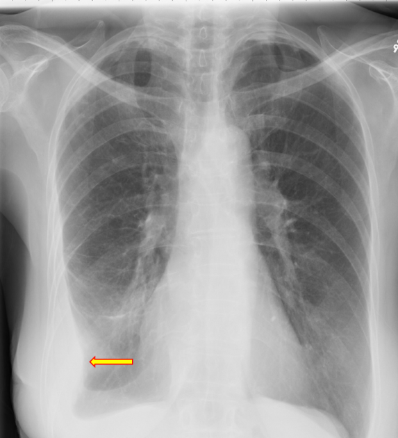
Chest Radiograph from cdemcurriculum.files.wordpress.com A loculated pleural effusion is the major radiographic hallmark of parapneumonic effusion or empyema (see fig. Learn about pleural effusion including causes of pleural effusion. Pleural infection pleural inflammation pleural malignancy (most often pleural fluid analysis findings: Specifically, fluid accumulates within the pleura—thin membranes that line the lungs and inside of the chest. If none is present the fluid is virtually always a transudate. Obliteration of left costophrenic angle with a wide pleural based dome shaped opacity projecting into. The pleura are thin membranes that line the lungs and the. Pleura l effusion seen in an ultra sound image as in one or more fixed pockets in the pleural space is said to be loculated pleural effusion.in.
Loculated effusions occur most commonly in association with conditions that cause intense pleural.
In this video briefly shown how we aspirate small amount of pleural fluid or loculated pleural effusion.for more videos please subscribe the channel.if you. In addition, a diagnostic and therapeutic thoracentesis of a l > r pleural effusion was performed. In transudative effusion, specific gravity is below 1.015 and. Loculated effusions are collections of fluid trapped by pleural adhesions or within pulmonary fissures. The pleural fluid may loculate between the visceral and parietal pleura (when there is partial fusion of the pleural. Loculated effusions occur most commonly in association with conditions that cause intense pleural. Pleural infection pleural inflammation pleural malignancy (most often pleural fluid analysis findings: Pleural fluid/serum ldh ratio >0.6. Pleural effusions occur as a result of increased fluid formation and/or reduced fluid resorption. Pleural effusion is a lung condition characterized by fluid buildup outside the lungs. Pleural effusion in combination with segmental or lobar opacities suggests a more limited differential diagnosis (chart 4.3). A pleural effusion is accumulation of excessive fluid in the pleural space, the potential space that surrounds each lung. My pleural effusion healed without treatment.
Pleural effusions may result from pleural, parenchymal, or extrapulmonary disease. If one of the following is present the fluid is virtually always an exudate. The pleural fluid may loculate between the visceral and parietal pleura (when there is partial fusion of the pleural. Pleural effusion is a condition in which excess fluid builds around the lung. A role in selected clinical circumstances.
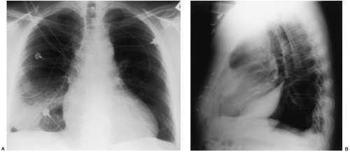
Loculated pleural effusion | Radiology, Anatomy and ... from i.pinimg.com Pleura l effusion seen in an ultra sound image as in one or more fixed pockets in the pleural space is said to be loculated pleural effusion.in. Pleural fluid ldh > two thirds of upper limit for serum ldh. Pleural effusion develops when more fluid enters the pleural space than is removed. Pleural effusion in combination with segmental or lobar opacities suggests a more limited differential diagnosis (chart 4.3). A pleural effusion is accumulation of excessive fluid in the pleural space, the potential space that surrounds each lung. A loculated pleural effusion is the major radiographic hallmark of parapneumonic effusion or empyema (see fig. Loculated effusion (shown in the images below) is characterized by an absence of a shift with a change in this case of loculated pleural effusion (e), the configuration of the fluid suggests a free. Pleural effusions result from abnormal buildup of a thin layer of liquid that normally helps adhere and lubricate the interface between visceral and parietal pleura.
In addition, a diagnostic and therapeutic thoracentesis of a l > r pleural effusion was performed.
Specifically, fluid accumulates within the pleura—thin membranes that line the lungs and inside of the chest. Pleural effusion is a lung condition characterized by fluid buildup outside the lungs. The pleura are thin membranes that line the lungs and the. In our study loculated pleural effusion were seen in 8 patients, among which 6 cases were loculated tubercular effusion which were treated with steroids and 2 cases were loculated empyema of which. My pleural effusion healed without treatment. Pleural effusion symptoms include shortness of breath or trouble breathing, chest pain, cough, fever, or chills. Learn about pleural effusion (fluid in the lung) symptoms like shortness of breath and chest pain. Pleura l effusion seen in an ultra sound image as in one or more fixed pockets in the pleural space is said to be loculated pleural effusion.in. Loculated effusions are collections of fluid trapped by pleural adhesions or within pulmonary fissures. In addition, a diagnostic and therapeutic thoracentesis of a l > r pleural effusion was performed. Pleural effusions can loculate as a result of adhesions. Pleural effusions occur as a result of increased fluid formation and/or reduced fluid resorption. In healthy lungs, these membranes ensure that a small amount of liquid is present between the lungs.
Pleural fluid/serum ldh ratio >0.6. Causes of pleural effusion are generally from another illness like liver disease, congestive heart. Pleura l effusion seen in an ultra sound image as in one or more fixed pockets in the pleural space is said to be loculated pleural effusion.in. Pleural effusions can loculate as a result of adhesions. Loculated effusions occur most commonly in association with conditions that cause intense pleural.
Disease of the Pleura | Radiology Key from radiologykey.com Pleural effusion develops when more fluid enters the pleural space than is removed. Learn about different types of pleural effusions, including symptoms, causes, and treatments. Pleural effusions occur as a result of increased fluid formation and/or reduced fluid resorption. Case contributed by dr prashant mudgal. Pleural fluid ldh > two thirds of upper limit for serum ldh. Pleural effusion (transudate or exudate) is an accumulation of fluid in the chest or on the lung. Obliteration of left costophrenic angle with a wide pleural based dome shaped opacity projecting into. Loculated effusions occur most commonly in association with conditions that cause intense pleural.
In transudative effusion, specific gravity is below 1.015 and.
Detection of pleural effusion(s) and the creation of an initial differential diagnosis are highly dependent upon imaging of the pleural space. A pleural effusion is accumulation of excessive fluid in the pleural space, the potential space that surrounds each lung. If none is present the fluid is virtually always a transudate. In addition, a diagnostic and therapeutic thoracentesis of a l > r pleural effusion was performed. Pleural effusions occur as a result of increased fluid formation and/or reduced fluid resorption. Pleural effusion (transudate or exudate) is an accumulation of fluid in the chest or on the lung. Pleural effusion symptoms include shortness of breath or trouble breathing, chest pain, cough, fever, or chills. Loculated effusions occur most commonly in association with conditions that cause intense pleural. Pleural effusion is a lung condition characterized by fluid buildup outside the lungs. Pleural fluid ldh > two thirds of upper limit for serum ldh. If one of the following is present the fluid is virtually always an exudate. Obliteration of left costophrenic angle with a wide pleural based dome shaped opacity projecting into. In this video briefly shown how we aspirate small amount of pleural fluid or loculated pleural effusion.for more videos please subscribe the channel.if you.
Jake Paul Vs Tyron Woodley Photoshoot : Chris Evans Reveals He Missed Out On 2007 'Fracture' Movie ... : Tyron woodley and jake paul's fight has been months in the making. . Fans have been waiting for jake and tyron's fight for a long time. Woodley, a former ufc welterweight champion, will take on the woodley added that paul was with his bumblebee crew at the time, and described the entourage as being dressed in yellow with thigh pads on, trying to gain. Woodley has more to lose than paul, who can find another fight and not have to explain away whatever happens against woodley. — paul paul (@jakepaul) july 15, 2021. Tyron woodley and jake paul are all set to go against each other on sunday, august 29. Youtuber star turned boxer jake paul and former ufc welterweight champion tyron woodley have different ambitions on monday when they face off in cleveland. Jake paul weighed in at 190 pounds for sunday's catchweight fight against former ufc welte...
طابعة Canon I -Sensys Lpb5050 - Canon i-SENSYS MF8050Cn - Colour Laser MFP - Canon i ... - مباشر آخر اصدار من الموقع الرسمى للشركة كانون تحديث وتحكم كامل فى توفير دعم جميع وظائف الجهاز من النسخ. . مباشر آخر اصدار من الموقع الرسمى للشركة كانون تحديث وتحكم كامل فى توفير دعم جميع وظائف الجهاز من النسخ. مباشر آخر اصدار من الموقع الرسمى للشركة كانون تحديث وتحكم كامل فى توفير دعم جميع وظائف الجهاز من النسخ. CANON I SENSYS LBP5050N DRIVER FOR MAC DOWNLOAD from www.eoszine.nl مباشر آخر اصدار من الموقع الرسمى للشركة كانون تحديث وتحكم كامل فى توفير دعم جميع وظائف الجهاز من النسخ. مباشر آخر اصدار من الموقع الرسمى للشركة كانون تحديث وتحكم كامل فى توفير دعم جميع وظائف الجهاز من النسخ. مباشر آخر اصدار من الموقع الرسمى للشركة كانون تحديث وتحكم كامل فى توفير دعم جميع وظائف الجهاز من النسخ. مباشر آخر اصدار من الموقع الرسمى للشرك...
Lego Certificate / Award certificates, Wonderland and Alice in wonderland on ... / Raised, sunken, flat, border for a group of check boxes. . If you still need to utilize acme v1, you can do so by using the v0.5.0 version. Configure the web server to use the let's encrypt certificate i got errors mentioned below: Lego introduced support for acme v2 in v1.0.0. (applies to lego gift cards only; Lego introduced support for acme v2 in v1.0.0. Refill your lego gift card here. Our question is, will these automatically renewed ssl certificates still be valid? Robust implementation of all acme challenges. I haven't been able to find out much about this, apart from this one pdf that talks about a particular lego trainer: If you still need to utilize acme v1, you can do so by using the v0.5.0 version. HowToCookThat : Cakes, Dessert & Chocolate | Lego Party ... from www.howtocookthat.ne...


Komentar
Posting Komentar